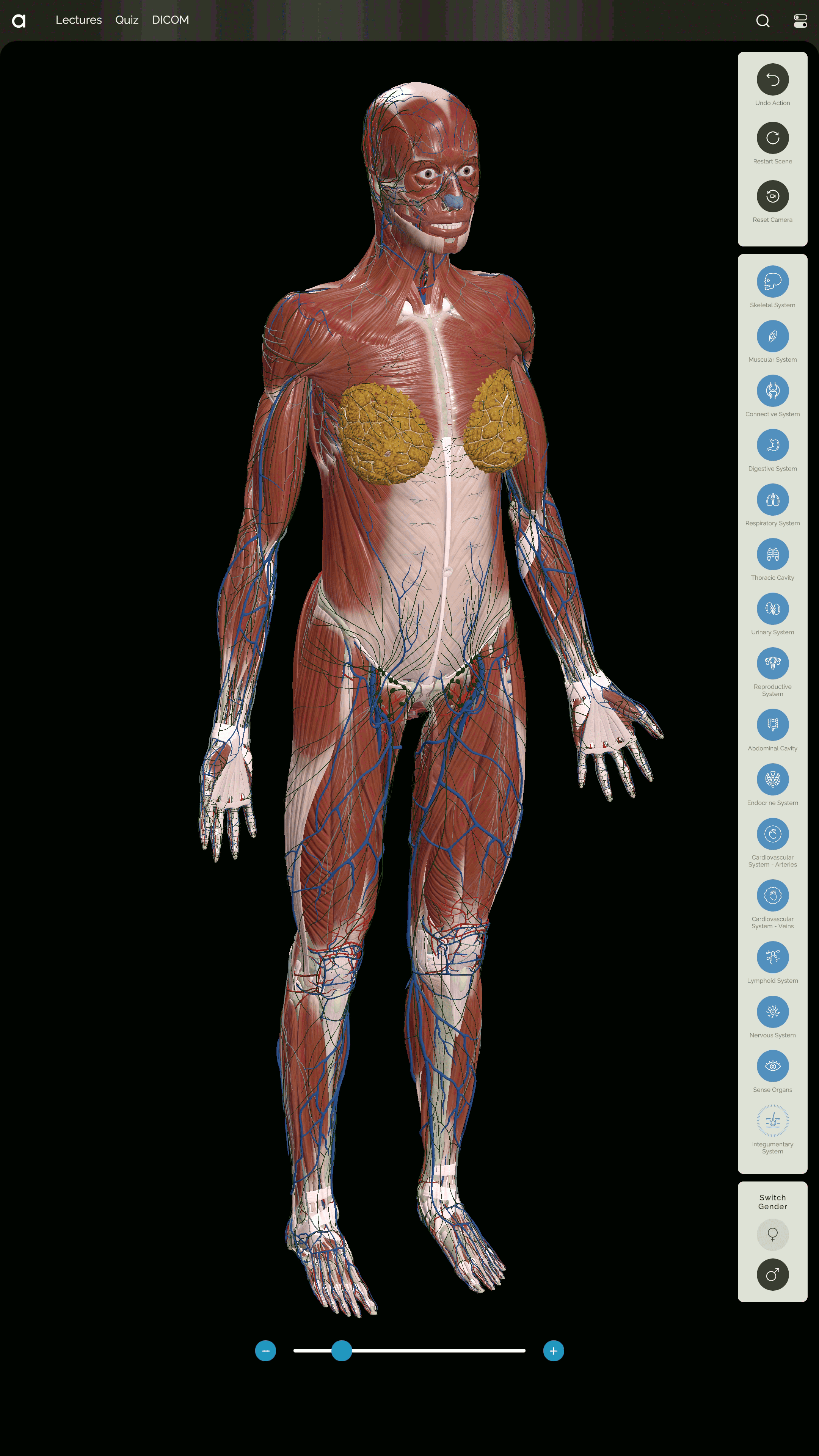
With the highest level of detail for the anatomical objects and structures of the human body for both sexes. Focus on each anatomical object and view its anatomical structures. Cross-sections of anatomical objects and cross-sectional views of all organs and systems are available. Dynamic search across all objects.
Innovative Electronic
Atlas of Human Anatomy
An advanced, AI-powered tool capable of visualizing and analyzing the anatomical structure of the human body and the results of medical research
Features
Anatomical atlas 
Quiz system

Allows you to not only study but also test your knowledge of human anatomy. It includes questions on both the names of anatomical objects and anatomical structures. Flexible testing options are available, including for specific human body system.
View DICOM
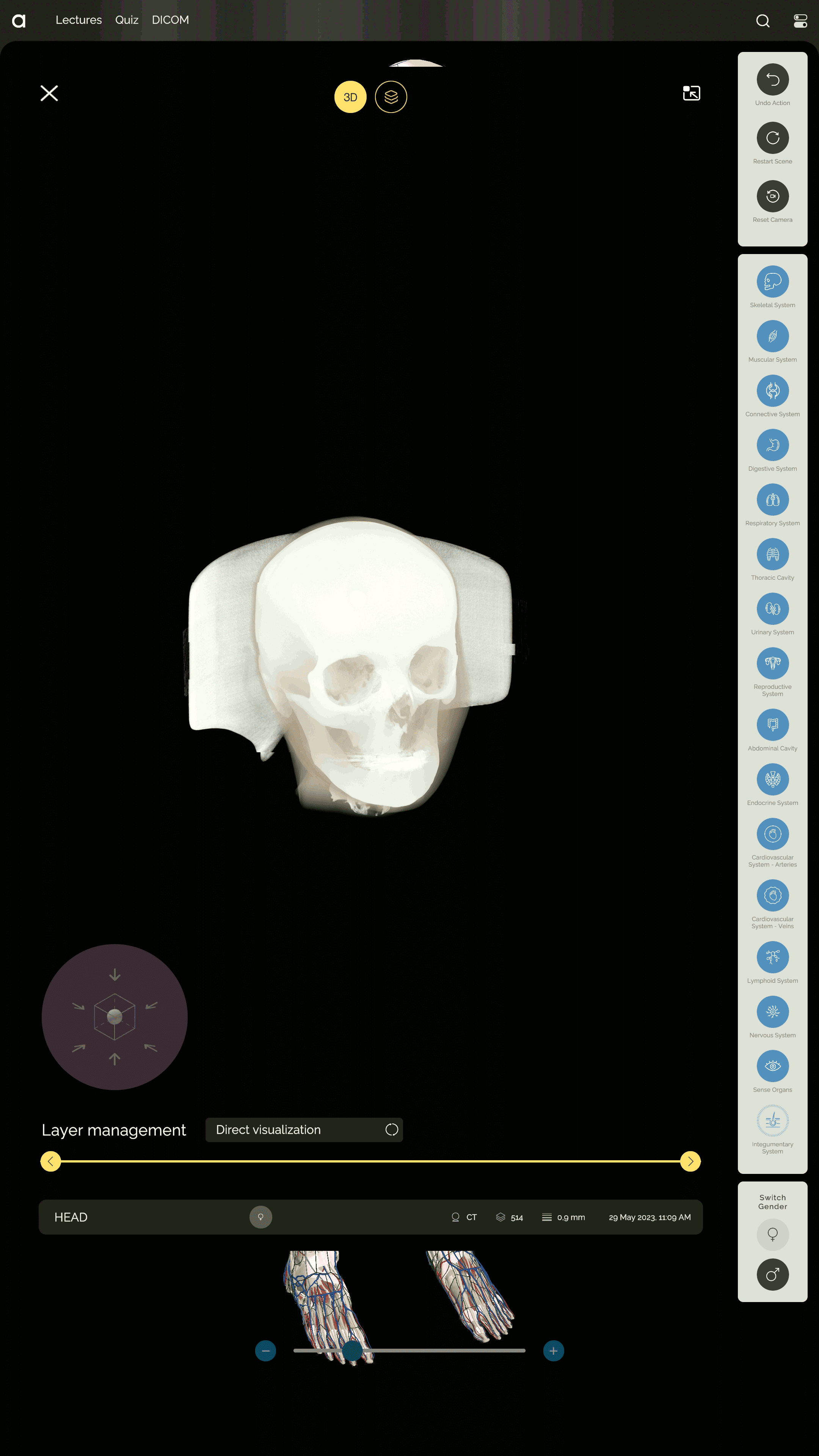
Studies on a large screen in a clear, easy-to-understand format for both physicians and patients. View image layers. Support for all study formats.
DICOM-based 3D model generation
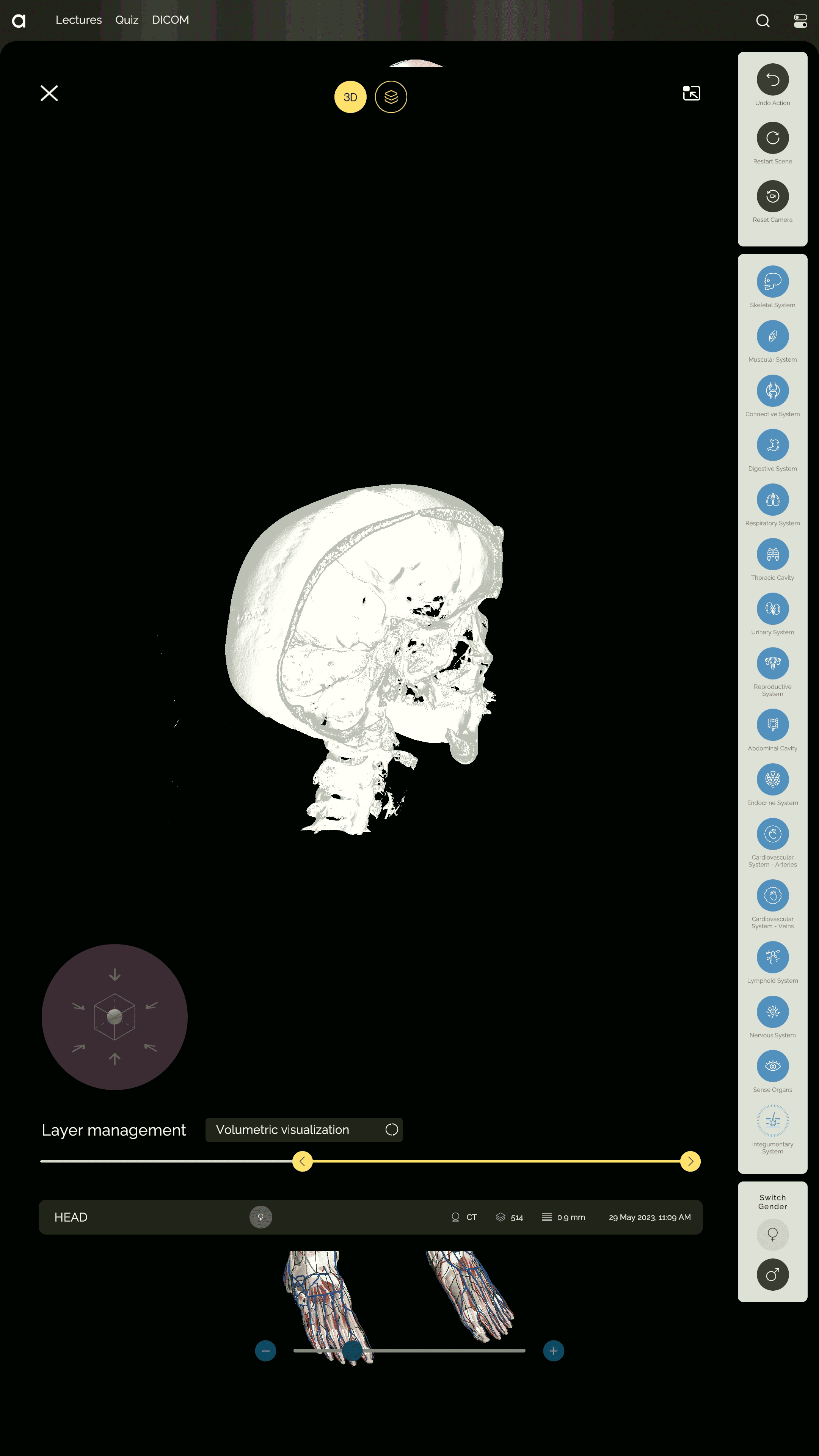
For detailed pathology diagnostics, dynamic sectioning, and tissue separation. Generation of a full-fledged 3D model based on multi-layer CT and MRI images. AI denoising of the model. Ability to quickly filter bone tissue. Dynamic tissue filtering on the 3D model
AI segmentation and analysis of DICOM
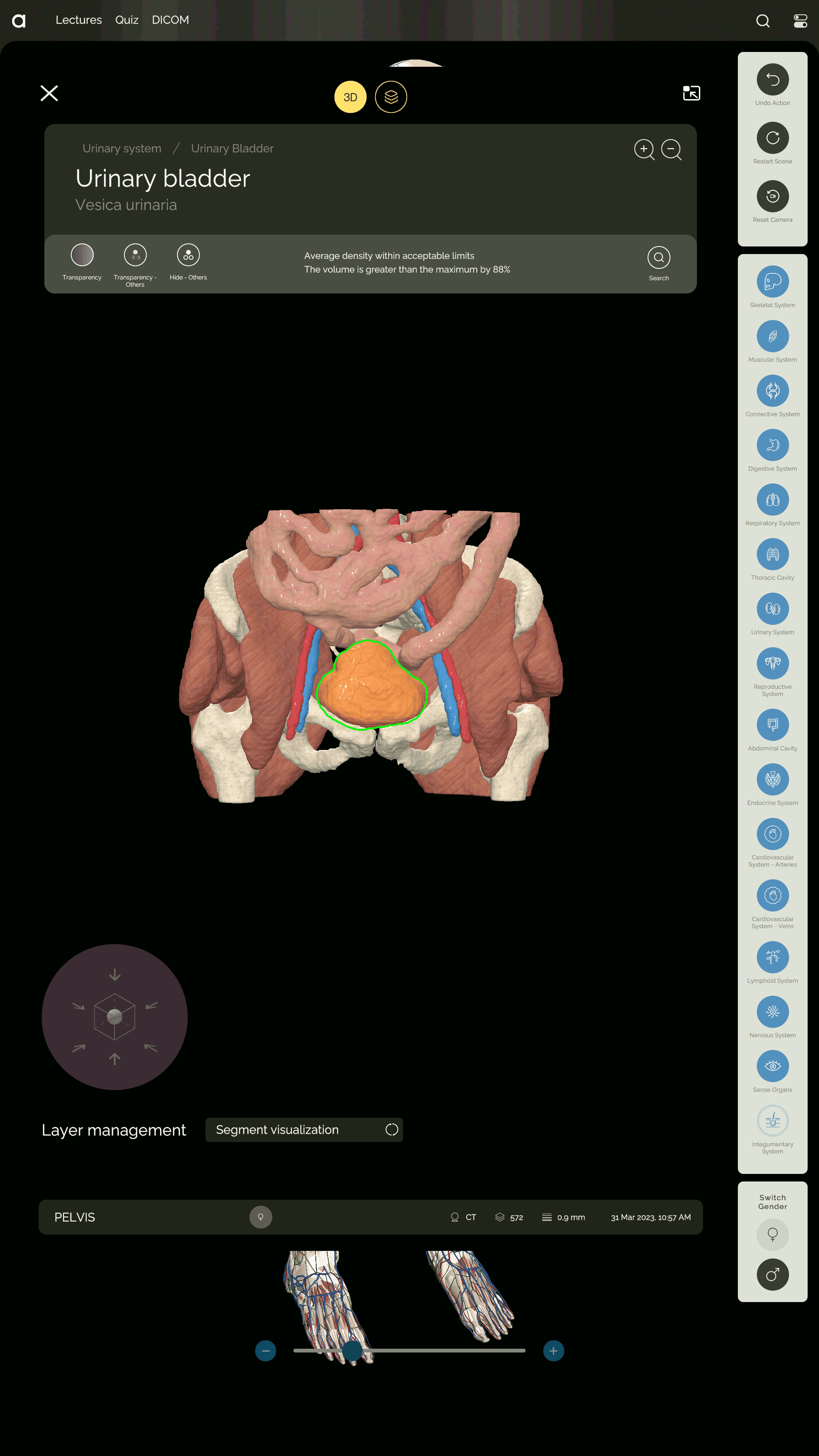
Segmentation of DICOM into separate anatomical objects using AI locally, while preserving the privacy of patient data. AI analytics and early diagnosis of pathological changes in the density and volume of anatomical objects in DICOM
4 500 +
Models of anatomical
objects
3 400 +
Anatomical
structures
1 500 +
Cross-sections
and constructs
10 000 +
Quiz questions

Compliance with the International Classification and Standard of Human Anatomical Nomenclature IFAA FIPAT

Developed with scientific support from leading anatomy experts
Anatomograph in education
One Anatomograph replaces more than 1000 anatomical posters, models and dummies
In the classroom
In the hall of the educational institution
Anatomograph in the classroom
Increasing engagement in the learning process
Highly visual and detailed models
An Anatomograph in the hall
of the educational institution
Working with an anatomical atlas outside of the classroom
Self-testing of knowledge in quiz mode
Anatomograph in practical medicine
Anatomograph is a unique device that makes diagnostics convenient and patient interactions visual through the use of AI
In the doctor's office
In the lobby of a medical facility
Anatomograph in the doctor's office
Analysis of test results using AI
Visual presentation of test results to the patient
Anatomograph in the lobby of a medical facility
Independent study of research results
Improving the status of a medical facility
Technical
characteristics
49"
high-definition touchscreen
Up to 10 simultaneous touches
Intuitive gesture control with on-screen gesture recognition for up to 10 simultaneous touches
8K
Connect an external monitor or projector with a resolution of up to 8K
Multilingual interface
Multilingual interface supporting English, Russian, Georgian, Arabic, and Latin
49’’
8K

Tilt up to 90°
Screen position control via remote control and buttons on the case: tilt up to 90°
Vandal-proof case
Rugged steel case
RGB backlight
RGB backlight control via remote control
Protection
4mm thick protective glass